Home

demonstration distance Human race immunofluorescence suspension cells Unpleasantly lack Partial

Coverslips for IF and suspension cells

How to Prepare your Specimen for Immunofluorescence Microscopy | Science  Lab | Leica Microsystems
How to Prepare your Specimen for Immunofluorescence Microscopy | Science Lab | Leica Microsystems

Immunofluorescence protocol for culture cells. EuroMAbNet

Protocol for adhesion and immunostaining of lymphocytes and other non-adherent  cells in culture | BioTechniques
Protocol for adhesion and immunostaining of lymphocytes and other non-adherent cells in culture | BioTechniques

Immunofluorescence Assays - Creative Bioarray

Suspension cell immunofluorescence

Microscopy
Microscopy

A) Immunofluorescence staining of SKBR3 E-cadherin and WT cells showing...  | Download Scientific Diagram
A) Immunofluorescence staining of SKBR3 E-cadherin and WT cells showing... | Download Scientific Diagram

Immunocytochemistry and immunofluorescence protocol | Abcam

Biology | Free Full-Text | Immuno-Electron and Confocal Laser Scanning  Microscopy of the Glycocalyx
Biology | Free Full-Text | Immuno-Electron and Confocal Laser Scanning Microscopy of the Glycocalyx

Shi-fix Coverslips – shikharbiotech.com

A sample preparation protocol for high throughput immunofluorescence of suspension  cells | bioRxiv
A sample preparation protocol for high throughput immunofluorescence of suspension cells | bioRxiv

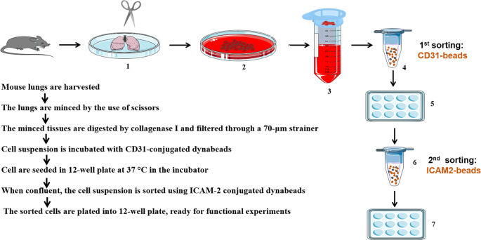
A simple protocol for isolating mouse lung endothelial cells | Scientific  Reports
A simple protocol for isolating mouse lung endothelial cells | Scientific Reports

Protocol for adhesion and immunostaining of lymphocytes and other non-adherent  cells in culture | BioTechniques
Protocol for adhesion and immunostaining of lymphocytes and other non-adherent cells in culture | BioTechniques

Adhesion of suspension cells on a coverslip in serum-free conditions -  ScienceDirect
Adhesion of suspension cells on a coverslip in serum-free conditions - ScienceDirect

Immunocytochemistry and immunofluorescence protocol | Abcam

Tips for Optimizing Immunofluorescence Protocols to Get the Best Image

Coverslips for IF and suspension cells

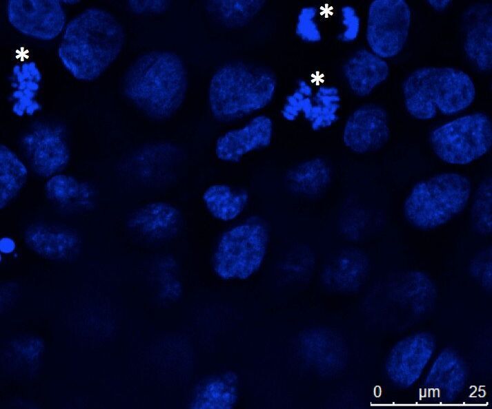
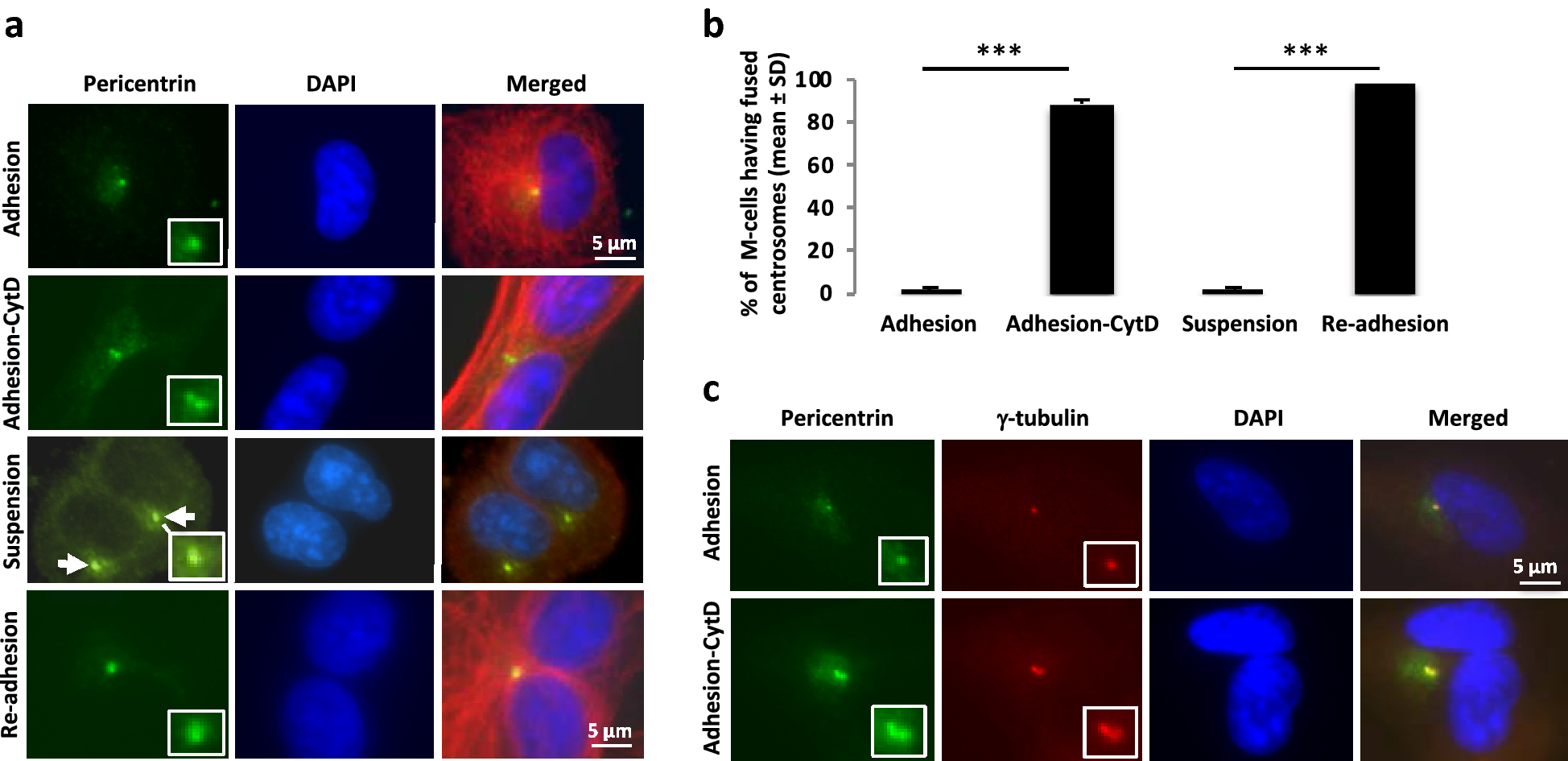
Septin and Ras regulate cytokinetic abscission in detached cells | Cell  Division | Full Text
Septin and Ras regulate cytokinetic abscission in detached cells | Cell Division | Full Text

Clusterin/APOJ Antibody (NBP1-06027): Novus Biologicals

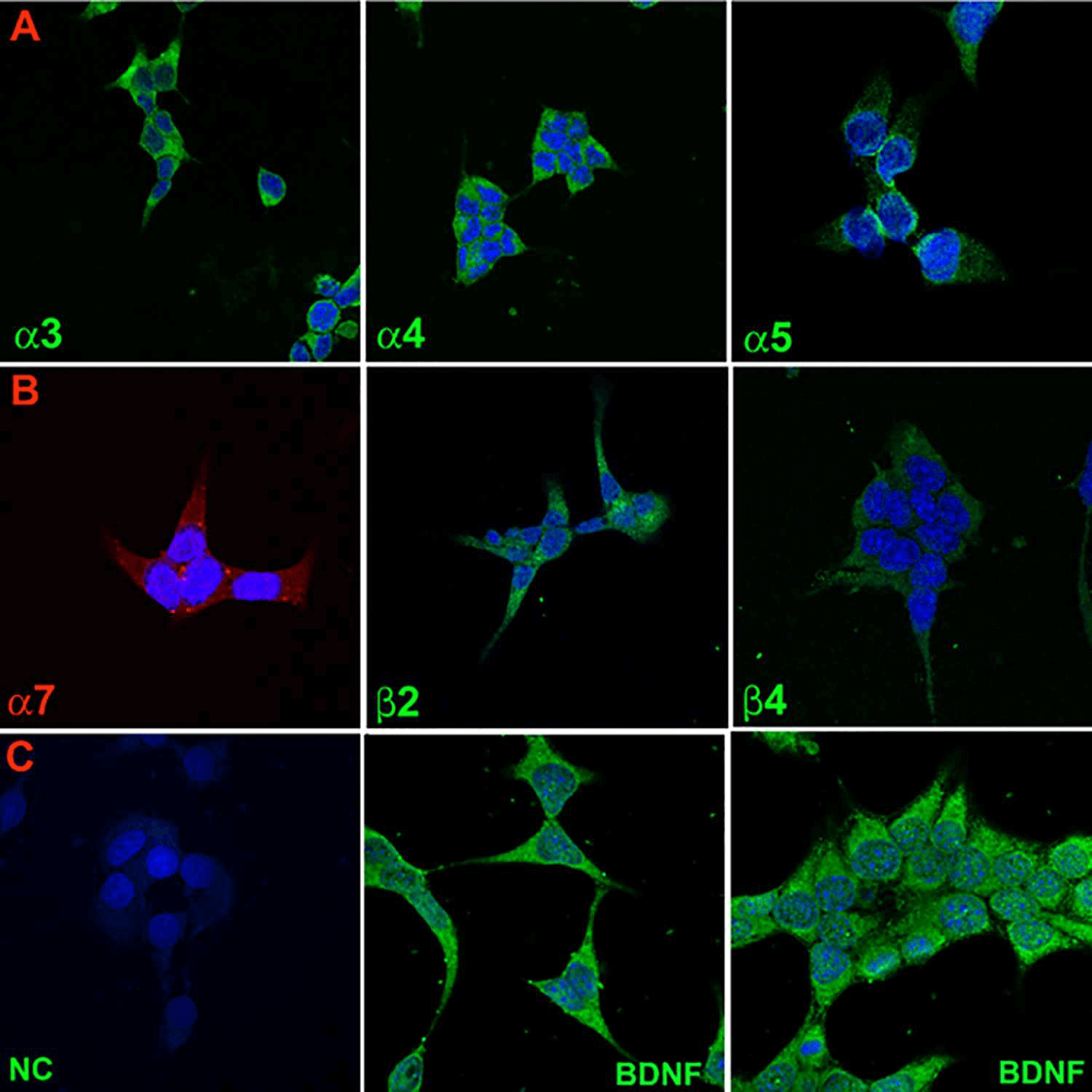
Immunofluorescence images illustrate that overexpressed, myc-tagged... |  Download Scientific Diagram
Immunofluorescence images illustrate that overexpressed, myc-tagged... | Download Scientific Diagram

Immunofluorescence staining for EMT markers. Suspension cells (NCI-H69)...  | Download Scientific Diagram
Immunofluorescence staining for EMT markers. Suspension cells (NCI-H69)... | Download Scientific Diagram

Tips for Optimizing Immunofluorescence Protocols to Get the Best Image

A Sample Preparation Protocol for High Throughput Immunofluorescence of Suspension  Cells on an Adherent Surface - Anna Bäckström, Laura Kugel, Christian  Gnann, Hao Xu, Joseph E. Aslan, Emma Lundberg, Charlotte Stadler, 2020
A Sample Preparation Protocol for High Throughput Immunofluorescence of Suspension Cells on an Adherent Surface - Anna Bäckström, Laura Kugel, Christian Gnann, Hao Xu, Joseph E. Aslan, Emma Lundberg, Charlotte Stadler, 2020

A sample preparation protocol for high throughput immunofluorescence of suspension  cells | bioRxiv
A sample preparation protocol for high throughput immunofluorescence of suspension cells | bioRxiv